Science Highlights, February 5, 2014
Awards and Recognition


Capability Enhancement
Improving plutonium-238 analytical chemistry operations
Awards and Recognition
Escobedo-Diaz, Fensin, Gibbs, Li, and Morrow receive Young Leaders Awards
The Minerals, Metals & Materials Society (TMS) has chosen five Los Alamos scientists to receive 2014 TMS Young Leaders Professional Development Awards. Juan (Pablo) Escobedo-Diaz (formerly of Materials Science in Radiation and Dynamics Extremes, MST-8), Saryu Fensin (MST-8), Paul Gibbs (Metallurgy, MST-6), Nan Li (Center for Integrated Nanotechnologies, MPA-CINT), and Benjamin Morrow (MST-8) will be honored in February during the TMS 2014 Annual Meeting and Exposition in San Diego, CA. Established to enhance the professional development of young researchers from TMS’s five technical divisions, the annual award opens opportunities to participate in Society activities, attend TMS conferences, network with Society members and leaders, receive mentoring from TMS division leaders, and serve as judges for division-sponsored student events at the TMS annual meeting.

Escobedo-Diaz
The Structural Materials Division nominated Escobedo-Diaz. He holds a PhD in materials science and engineering from Washington State University. Following a postdoctoral position at its Institute of Shock Physics, he joined MST-8 as a postdoctoral researcher. Ellen Cerreta (MST-8) and Darcie Dennis-Koller (Shock and Detonation Physics, WX-9) mentored him. He spent three years at LANL focused on understanding the linkages between microstructure of metals and dynamic damage evolution. Laboratory Directed Research and Development (LDRD) sponsored the work, which led to the development of damage studies that NNSA Science Campaign 2 funded. Escobedo-Diaz’s portion of the research has resulted in more than 10 peer-reviewed journal papers. He has joined the faculty of the University of New South Wales, Australia.

Fensin
Fensin received the award in the Electronic, Magnetic & Photonic Materials Division. She received a PhD in materials science and engineering from the University of California, Davis, and then joined MST-8 as a postdoctoral researcher in 2010. Ellen Cerreta, Steve Valone, and Rusty Gray (MST-8) mentored her. Fensin is an expert in molecular dynamics simulations and experiments involving interfaces in mechanical and thermodynamic extremes. She studies the behavior of material microstructure (single and bi-metal interfaces) on damage nucleation and evolution under dynamic loading conditions. As a staff member, her work is funded through both DOE Office of Basic Energy Sciences Energy Frontiers Research Center –The Center for Materials in Irradiation and Mechanical Extremes (CMIME) and a NNSA Science Campaign 2 project to examine damage in two-phase metals. She serves on the TMS committees of Mechanical Behavior of Materials, Young Professionals, and Chemistry and Physics of Materials committees.

Gibbs
The Materials Processing & Manufacturing Division nominated Gibbs. He received a PhD in metallurgical and materials engineering from the Colorado School of Mines in 2012. Gibbs is a Metallurgy (MST-6) postdoctoral researcher working with Amy Clarke (MST-6) to examine the influence of imposed processing variations on solidification microstructure evolution using real-time X-ray and proton imaging. Amy Clarke's Early Career award from the DOE, Office of Basic Energy Sciences, Division of Materials Sciences and Engineering funds his research. He received a 2013 American Iron and Steel Institute Finalist Medal for a paper describing a methodology to produce a family of transformation-induced plasticity steels with desirable austenite stability conditions.
Li
The Materials Processing and Manufacturing Division nominated Li. He earned a PhD in materials science at Texas A&M University. Li joined CINT to work with Amit Misra (MPA-CINT) as a graduate research assistant and then as a postdoctoral researcher. He became a staff scientist in MPA-CINT in 2013. Through a combination of electron tomography and in situ transmission electron microscopy, Li aims to understand how atomic structures of interfaces contribute to the evolution of deformation-induced or radiation-induced defects and damage in advanced materials. The DOE, Office of Science, Office of Basic Energy Sciences sponsors the work. He has more than 30 peer-reviewed publications (17 first-authored), two of which were listed in the ScienceDirect TOP25 Hottest Articles in Materials Science. He has been awarded TMS Best Graduate Student Paper Award, ACTA Student Award, and the LANL Distinguished Postdoctoral Performance Award.

Morrow
Morrow received the award from the Structural Materials Division. He earned a PhD in materials science and engineering from The Ohio State University. As a postdoctoral researcher with Ellen Cerreta (MST-8), he focuses on characterization of materials and structure-property relationships in metals. Morrow conducts in situ experiments in a transmission electron microscope (TEM) to directly observe deformation mechanisms at the structure of twin boundaries. The DOE Office of Basic Energy Sciences funds the project to investigate the constitutive response of hexagonal metals, and a NNSA Science Campaign 2 project sponsors study of the role of dynamic drive condition on microstructural evolution in zirconium. He serves on the TMS committees Mechanical Behavior of Materials, Titanium, Young Professionals, and Women in Science. An honorable mention award at the Lab’s 2013 Postdoc Research Day recognized his work.
TMS is an international professional organization of more than 12,000 members, encompassing the entire range of materials and engineering, from minerals processing and primary metals production to basic research and the advanced applications of materials. Technical contacts: Ellen Cerreta and Amit Misra
Joe D. Thompson wins Frank H. Spedding Award
Joe D. Thompson (Condensed Matter and Magnet Science, MPA-CMMS) will receive the 2014 Frank H. Spedding Award “for outstanding contributions to the physics of f-element materials, especially their magnetism and unconventional superconductivity.” The award, which is given in recognition of excellence and achievement in research on the science and technology of rare earths, is named for Frank Spedding (1902-1984), a Canadian chemist who developed the Ames process to achieve high purity uranium for the Manhattan Project’s atomic bomb.
Joe D. Thompson
Thompson received a PhD in physics from the University of Cincinnati, and then joined the Lab as a postdoc in 1975. He is a Fellow of the American Association for the Advancement of Science, American Physical Society, and LANL. Los Alamos, DOE, and the Japan Society for the Promotion of Science have given him research awards. He is a member of the Institute for Scientific Information inaugural group of the top 150 most frequently cited physicists in the world.
Thompson will receive the award during the 27th Rare Earth Research Conference in Reno, NV. The meeting is the premier gathering for multidisciplinary basic and applied research on the f-elements. He will present an invited talk on his research at the conference. Technical contact: Joe Thompson
Biophysical Society profiles Sandrasegaram Gnanakaran
The January 2014 Biophysical Society newsletter contained a “Biophysicist in Profile” article regarding Sandrasegaram Gnanakaran, aka “Gnana,” of Theoretical Biology and Biophysics (T-6).
The article describes his childhood and education in Sri Lanka and how he became interested in research. Due to the violent civil war in Sri Lanka, Gnana came to the U.S. to continue his education in a more stable environment. His experiences as an undergraduate at Virginia Commonwealth University motivated him to pursue graduate studies in computer science and chemistry. He received a PhD in physical chemistry at the University of Pennsylvania, where he studied energy transfer and relaxation of vibrationally excited molecules in different solvents. Gnana became interested in research at the interface of biological and physical sciences and tackling a scientific problem from many different angles. As a postdoc at the University of Pennsylvania, Gnana conducted theoretical interpretation of the new 2D-IR spectroscopy. He used molecular dynamics simulations to deduce conformations of peptides in conjunction with the spectroscopic measurements.
Sandrasegaram Gnanakaran
Gnana joined LANL as a postdoc with Angel Garcia to apply theoretical approaches for the study of protein dynamics, folding, and misfolding. After completing his postdoc, he became a staff scientist.
Gnana takes an interdisciplinary approach for his research. He and Byron Goldstein (T-6) are building detailed models of cell signaling cascades. Gnana also leads a project to examine efflux pump mediated drug resistance, the dominant drug resistance mechanism in Gram-negative bacteria. The researchers are developing an experimentally-driven mathematical framework that will integrate structural, genetic, and cellular processes to understand how multi-drug resistance efflux pumps are able to resist so many antibiotics. In another research area, Gnana and his collaborators are establishing theoretical capabilities to overcome challenges related to cost-effective biofuels. In the case of cellulosic conversion of glucose to ethanol, they are examining how three kinds of enzymes work together in synergy to break down crystalline cellulose to glucose. LANL teams with the Great Lakes Bioenergy Center to identify which properties determine efficient catalysis of an enzyme on native and non-native cellulose surfaces. They have also developed mathematical models to identify the proper mixture of these enzymes that effectively degrade cellulose.
The Biophysical Society has more than 10,000 members who work at the interface of biology and physics. Technical contact: Sandrasegaram Gnanakaran
Capability Enhancement
Improving plutonium-238 analytical chemistry operations
The installation of DC Arc and plutonium Assay/Radiochemistry operations into TA-55/PF4 allows plutonium-238 (238Pu) operations to be removed from the CMR facility. The operations are a significant contribution to the Material at Risk (MAR) at CMR, a facility scheduled for closure in ~2019. Analytical chemistry of the 238Pu product is paramount in supporting the fuel requirements for NASA, NE-75, and DP customers. The transition of Pu-238 analytical chemistry to TA-55/PF4 has included the design, fabrication and installation of three new gloveboxes and development of methods and instrument technique. Management completed the self-assessment (MSA) for installation, and 238Pu operations are planned to begin in 2014. The team accomplished this work ahead of schedule and on or below budget.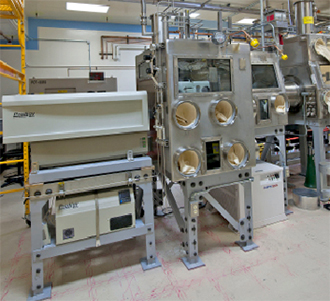
Photo. The DC Arc spectrometer installed in PF4 replaces an instrument that is over 40 years old.
DC arc capability. The team installed the DC Arc glovebox in PF4 room 201 and conducted a MSA. The DC Arc process is an optical emission technique that performs spectroscopic analysis of solid PuO2 sample material to determine elemental impurities. The researchers conducted method development to transition from a 40-year-old instrument that used photographic plates to the new, compact, faster, more streamlined instrument and process. The team conducted the work in conjunction with a supplier of inductively coupled plasma (ICP) instruments to incorporate technology from the spectrometer. The new DC Arc instrument builds upon the established ICP technology.
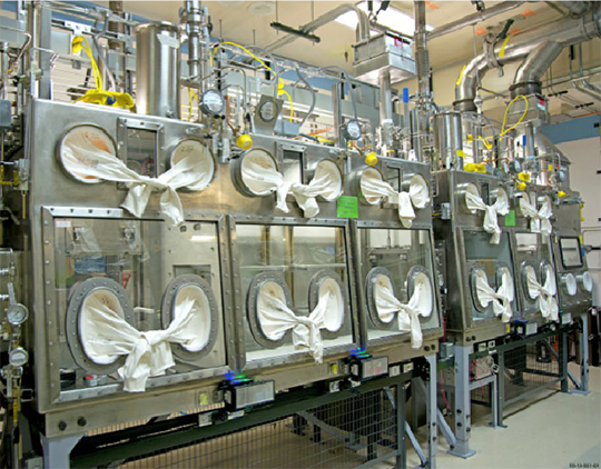
Photo. The new gloveboxes improve 238Pu analytical chemistry operations.
Plutonium assay and radiochemistry. The two gloveboxes installed for Pu assay and radiochemistry operations have many features that provide added safety and ergonomic design such as oval glove ports, large viewing windows, fire suppression, fire skirting, corrosion coatings, pneumatic actuated airlock doors, and more. As part of the transition, the team developed new separation methods to eliminate hazardous chemicals, as well as streamlining counting techniques. The researchers implemented a non-destructive Pu isotopic method using gamma counting with FRAM software in place of thermal ionization mass spectrometry (TIMS). The new method will reduce time spent in glovebox work (minimizing dose) as well as shortening turn-around time. The team developed a new separation process to prepare samples for TIMS analysis, which can be accepted within the MAR limits at the Radiological Laboratory/ Utility/Office Building (RLUOB). In addition to the ability to measure Pu isotopics non-destructively, workers have developed new non-destructive analysis methods for neptunium-237 (237Np), americium-241 (241Am), and plutonium-236 (236Pu) that will save time, cost, and reduce radiation dose to the workers.
NNSA funded the work, which supports the Lab’s Nuclear Deterrence mission area and the Materials for the Future and Science of Signatures science pillars. The team includes the Actinide Analytical Chemistry Group (C-AAC) and Nuclear and High Hazard Operations (NHHO). Technical contact: Nell Carver
Increased energy efficiency for computer data centers
The DOE has set a standard for all facilities such as LANL, Sandia National Laboratories, Oak Ridge National Laboratory, and Lawrence Livermore National Laboratory to operate at a Power Usage Effectiveness (PUE) ratio at or less than 1.4 in order to reduce energy consumption as well as operating costs. The PUE is a measurement of how efficiently a computer data center uses energy. The ratio is determined by dividing the total amount of energy a facility uses by the total amount of energy used by IT equipment and computer data centers.
Computer Room Air Conditioning (CRAC) units keep the cluster hardware located in the data centers cool. Prior to this project, units were set to supply a temperature of 55 degrees Fahrenheit to the computer data centers. Developments in computing architectures have now made it possible to supply clusters with warmer air from CRAC units without damaging cluster hardware.
In April of 2013, High Performance Computing (HPC) Division and Computer Computational and Statistical Sciences (CCS) Division began a collaboration to find the optimal room temperature at which CRAC units could supply cool enough air for the clusters and also run more efficiently. The team collected baseline data from the clusters, CRAC units, and temperatures around the room through an Environet application that uses wireless sensors placed around the computer rooms. This information helped establish the protocols to apply optimal conditions on computing room floors at LANL.
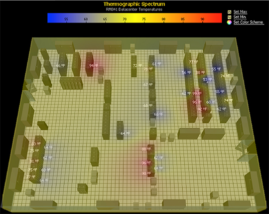
Figure 2. Thermographics of LDCC Computer Room 341 as viewed from Environet.
Several conditions had to be considered for this process. Each cluster has a thermal “sweet-spot” that makes the cluster function efficiently with a mix of individual computing nodes, or networked computers, running between an idle state at a lower temperature and a utilized state that could be considerably hotter when running a particularly data intensive data set. The optimal operating temperature of a node’s Central Processing Unit (CPU) had not been established. The team gathered specifications from the manufacturers for the upper operating temperature limit. The team also considered that each room contains a variety of computing architectures and equipment for the computing clusters, as well as storage tape silos, support servers, and network interconnects for future projects.
The team selected room 341 in the Laboratory Data Communications Center (LDCC) to study first. Room 341 currently houses Moonlight, Mustang, and Pinto for the Institutional Computing (IC) and Advanced Simulation and Computing (ASC) programs. Other applications such as Tivoli Storage Manager (TSM), file systems, networking and support services for HPC use are also housed in the room. The room 341 baseline data revealed that the room was substantially overcooled.
The team selected room 341 in the Laboratory Data Communications Center (LDCC) to study first. Room 341 currently houses Moonlight, Mustang, and Pinto for the Institutional Computing (IC) and Advanced Simulation and Computing (ASC) programs. Other applications such as Tivoli Storage Manager (TSM), file systems, networking and support services for HPC use are also housed in the room. The room 341 baseline data revealed that the room was substantially overcooled.
New instrument aids fission-fragment yield measurements
Uranium undergoes fission with thermal neutrons. The fissioning isotope, uranium-235 (235U), splits into two asymmetric fragments. The distribution of the mass of these fragments, the mass yield, is an important test for different theoretical models of nuclear fission. It also provides important data to nuclear weapons diagnostics, enables determination of the burn-up of nuclear fuel, and calculation of the source term for spent fuel waste stream analysis. Future measurements of plutonium-239 (239Pu) fission yields will provide a definitive answer regarding the energy dependence of neodymium-147 (147Nd), which is of importance to the weapons program.
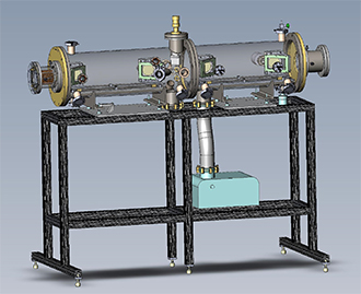
Figure 3. Illustration of the SPIDER spectrometer without its case. The source is in the middle. Time pickoff detectors, which use an electrostatic field to focus electrons from a foil to a microchannel-plate detector, are evident on both sides of the fissile foil in the middle.
The Nuclear Science group (LANSCE-NS) developed an instrument to measure fission-fragment yields as a function of the incoming neutron energy, the fragment mass and charge, and the total kinetic energy of the fragments. The team designed the instrument, named SPIDER (SPectrometer for Ion DEtermination in fission Research), to achieve 2 - 5% accuracy for incoming neutrons between 0.01 eV - 20 MeV. The scientists use the “2E-2V” method to measure the masses of the fragments. The energy of each fragment is determined by 1) its energy deposition in an ion-chamber and 2) its velocity by time-of-flight. This provides a mass resolution of better than 1 amu and approximately 1 charge unit for the light fragment.
Further work is needed before drawing conclusions about the agreement between the evaluated data (ENDDF/B-VII.1) and this experiment. The team will perform an improved calibration of the instrument using radiation sources at the end of the current LANSCE run cycle. Future plans include the construction of 8-10 detector arms to enhance the acquisition rate and enable the measurement of fast-neutron-induced yields.
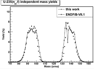
Figure 4. Preliminary results from the measurement performed at the Lujan Center at LANSCE.
A collaboration between Los Alamos, University of New Mexico, Colorado School of Mines, Slovak Academy of Sciences, Lawrence Livermore National Laboratory, and Lawrence Berkeley National Laboratory enabled detector design, construction, and analysis. The LANL team included Fredrik Tovesson, Krista Meierbachtol, and Dan Shields (LANSCE-NS); Morgan White (Materials and Physics Date, XCP-5), Charles Arnold and Todd Bredeweg (Nuclear and Radiochemistry, C-NR); Justin Jorgenson (Applied Engineering Technology, AET-5); and Arnie Sierk (Nuclear and Particle Physics, Astrophysics and Cosmology, T-2).
The Laboratory Directed Research and Development (LDRD) Program funded the instrument’s construction and research, which supports the Laboratory’s Nuclear Deterrence and Global Security mission areas and the Nuclear and Particle Futures science pillar. Technical contact: Fredrik Tovesson
Earth and Environmental Sciences
Making decisions about contaminated groundwater
A scientifically defensible decision process requires consideration of the uncertainties involved. Decisions related to subsurface contaminant remediation are often made with severe uncertainties that create challenges for applying traditional Bayesian (probability-based) approaches. Uncertainties in naturally occurring processes and remedial activities impact predictions of future contaminant concentrations. Therefore, it is prudent to incorporate these uncertainties into the decision process when designing or deciding between remediation methods within budget constraints. D. O'Malley and V. V. Vesselinov (Computational Earth Science Group, EES-16) developed an approach based on the Information Gap Decision Theory (IGDT) that accounts for lack of knowledge in naturally occurring dispersion and the functional form of the transport model. The IGDT approach is non-probabilistic and requires neither prior nor posterior knowledge about the frequency of occurrence of possible outcomes. The journal Water Resources Research published the research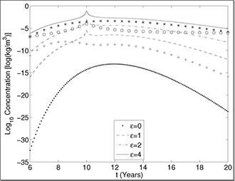
Figure 5. The plot depicts the maximum possible contaminant concentrations (dotted and dashed lines) at different levels of uncertainty, epsilon, as well as the actual concentrations (*, O, +) for three plausible contaminant transport scenarios. Predicted concentrations for enhanced dispersion (*), heavy-tailed dispersion (O), and a model with a secondary (earlier time arriving and less massive) source (+).
In the context of contaminant remediation, the IGDT approach examines the worst-case scenario as a function of the unknown horizon of uncertainty. The scientists applied this approach to two different scenarios representing typical groundwater contamination problems. The two remediation choices are to either remove contaminant mass directly, or increase the reaction rate. In one scenario, a remedial approach based on artificially increasing a reaction rate that reduces contaminant concentrations was preferred. This was due in large part to the fact that 10 years elapse before the peak contaminant concentrations are observed at the point of compliance measurement. This provided sufficient time for the increased reaction rate to reduce the contaminant concentration drastically. In the other scenario, only one year elapses before the peak contaminant concentrations are observed at the point of compliance, and the preferred remediation method depended on the remediation budget. These scenarios demonstrate that IGDT provides a viable tool for supporting decisions related to contaminant remediation by taking into account uncertainties and implementation costs. IGDT analyses are also applicable to other environmental problems such as carbon dioxide sequestration and hydro fracturing as well as to national security decision issues such as nuclear waste deposition, threat reduction, nuclear test deterrence, and stockpile stewardship.
Reference: “Groundwater Remediation Using the Information Gap Decision Theory,” Water Resources Research 50, 1 (2014); doi: 10.1002/2013WR014718, 2014.
The LANL Environmental Programs Directorate, DOE Advanced Simulation Capability for Environmental Management (ASCEM) project, DOE, Environmental Management; and the Integrated Multifaceted Approach to Mathematics at the Interfaces of Data, Models, and Decisions (DiaMonD) project, DOE, Office of Science funded different aspects of the work. The research supports the Laboratory mission areas and the Information, Science, and Technology science pillar. Technical contacts: Daniel O'Malley and Monty Vesselinov
Materials Physics and Applications
Coherent diffractive imaging featured in JOM and in symposium
The September 2013 issue of JOM, the TMS (The Minerals, Metals & Materials Society) member journal, featured a special section on high-resolution X-ray coherent diffractive imaging (CDI) edited by John Carpenter (Metallurgy, MST-6) and Richard Sandberg (Center for Integrated Nanotechnologies, MPA-CINT). Although the concept of coherent diffraction imaging has been around since the 1950s, experimental realization of this technique occurred only about fifteen years ago. CDI has produced high-resolution images from diffraction patterns of both crystalline and non-crystalline materials and has allowed imaging and characterization to occur for a broad range of materials. Unique characterization abilities such as three-dimensional strain mapping and non-destructive three-dimensional tomographic imaging highlight the emerging importance of CDI in the field of materials science. The series of three invited papers examined current and potential applications of CDI techniques to solve problems in materials science. The applications offer a way to foster increased interaction between physicists (the predominant purveyors of this technique) and materials scientists (potential customers).
References: “Perspective on the Use of Coherent Diffraction Imaging as a Tool for High Resolution Materials Characterization,” JOM 65, (9) 1181 (2013); doi: 10.1007/s11837-013-0671-7. Authors include John Carpenter (MST-6) and Richard Sandberg (MPA-CINT).
“Studies of Materials at the Nanometer Scale Using Coherent X-Ray Diffraction Imaging,” JOM 65, (9) 1208 (2013); doi: 10.1007/s11837-013-0699-8. Authors include Ricahrd L. Sandberg (MPA-CINT), Zhifeng Huang, Rui Xu, and Jianwei Miao (University of California, Los Angeles); and Jose A. Rodriguez
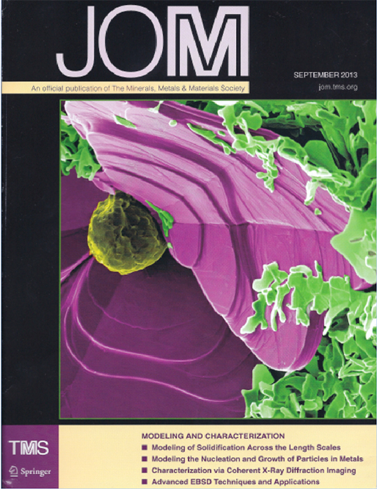
Figure 6. Cover of the journal.
In conjunction with this special section in JOM, Ross Harder (Argonne National Laboratory), Sandberg, and Carpenter organized a new symposium, “Characterization of Materials through High Resolution Coherent Imaging,” at the 2013 TMS Annual Meeting. The symposium supported the JOM special section editors’ desire to create more interaction between the CDI field and the greater materials science community. Researchers presented 13 invited talks and 10 submitted talks, including talks from Sandberg, Cris Barnes (Physics, P-DO), George Havrilla (Chemical Diagnostics and Engineering, C-CDE), as well as many coauthored presentations from LANL.
Invited presentations from Los Alamos researchers include:
Response to the new symposium from the speakers, of whom a majority were physicists, and the predominantly materials science-focused audience was overwhelmingly positive. The editors hope that this JOM section leads to collaborations that can be presented in another CDI symposium at the TMS Annual Meeting in 2015 in Orlando, FL. Technical contact: Richard Sandberg
Materials Science and Technology
Proton radiography probes metal solidification
For the first time, high-energy protons have been used to image a large metal sample during melting and solidification without destroying the sample. Such real-time imaging will provide the insights needed to control the microstructure of metals and lead to advanced manufacturing processes that fabricate materials with desired properties. Creation of microstructures by design is considered a grand challenge in this field. Scientific Reports published the work, and the DOE, Office of Science, Basic Energy Sciences website highlighted the paper: http://science.energy.gov/bes/highlights/2013/bes-2013-06-a/
Invented at Los Alamos, modern proton radiography (pRad) penetrates materials with a high-energy proton beam to produce pictures and time-lapse movies of the properties and behavior of materials under a variety of conditions and three-dimensional processes. This method has several advantages compared with X-ray radiography. X-rays are typically used on low-density metals of small volumes (<1 mm3), whereas proton radiography can be used on large samples (>10,000 mm3). The increase of more than four orders of magnitude in sample size for pRad enables more realistic observations of the metal solidification processes.
In this work, researchers used 800 MeV protons at the Los Alamos Neutron Science Center (LANSCE) and synchrotron X-ray radiography at Argonne National Laboratory’s Advanced Photon Source to interrogate solidification in an alloy of aluminum with 10 atomic percent indium additions. This alloy exists as two separate liquid phases before solidification. The scientists monitored the fluid flow and structure evolution in the experiments. The team made the first-ever pRad video sequence of aluminum with 10 atomic percent indium melting and solidification. The minority and majority liquid phases behave similarly to oil droplets in water, except that the denser, indium-rich liquid droplets sink instead of floating in the majority aluminum-rich liquid phase. Understanding the complex motion and coarsening of the indium-rich liquid droplets in the melt may provide a methodology to control how the solidification structure develops and allow improved casting quality.
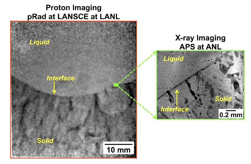
Figure 7. Protons and X-rays permit nondestructive and direct imaging of metal alloy melting and solidification. The left image demonstrates that protons can be used to examine a large sample volume during solidification, which is relevant for casting. The right inset image highlights a representative sample volume probed using X-rays. The images depict solidification structure development in an aluminum – 10 atomic percent indium alloy at different length scales.
Manipulation of solidification parameters is crucial for growing single crystals or controlling structure evolution and micro-segregation in a casting, setting the stage for what properties are possible after any downstream processing. Large scale parameters are controlled by casting design, which can include highly engineered melt flow patterns, heat transfer during mold filling, and progression of the solidification reaction. This early processing path determines the overall quality of a casting by influencing the presence of defects and the microscopic phase arrangement – characteristics that persist even after subsequent thermal-mechanical processing. Current methods to design castings incorporate experimental trials with destructive characterization techniques and empirical modeling. Post-mortem structure evaluations are typically used to infer what occurred at elevated temperatures, but direct observations of metallic alloy solidification have been limited. Commercially available tools to simulate fluid flow and solidification sequence are generally finite-element based and rely largely on post-processing and empirical relationships to simulate structural outcomes.
Proton microscopy fills a critical gap in the evolving capabilities of dynamic imaging techniques. It enables direct observations of structural outcomes as a function of processing in large volumes of materials. It also affords studies of three-dimensional processes, such as fluid flow encountered during solidification for which thick sections, rather than thin (constrained) sections, better represent processes that occur in actual castings. The real-time monitoring of metallic alloys during processing will permit direct interrogation of responses to parameter changes needed to develop predictive structure evolution models to couple with finite elements. This information will bridge the micro- and macro-scale regimes. It will also enable directed synthesis and processing to control structure evolution and the creation of optimal properties during process development.
Reference: “Proton Radiography Peers into Metal Solidification,” Scientific Reports 3, 2020 (2013); doi: 10.1038/srep02020. Los Alamos contributors include principal investigator Amy Clarke, Seth Imhoff, Paul Gibbs, Jason Cooley, Tim Tucker, Kester Clarke, Joel Montalvo, Robert Field, James L. Smith, Thomas Ott, and Martha Barker (Metallurgy, MST-6); Christopher Morris, Brian Hollander, and Fesseha Mariam (Subatomic Physics, P-25); Frank Merrill (Neutron Science and Technology, P-23); Brian Patterson (Polymers and Coatings, MST-7); Dan Thoma, and David Teter (Materials Science and Technology, MST-DO). Collaborators are Wah-Keat Lee (Brookhaven National Laboratory), Kamel Fezzaa, and Alex Deriy (Argonne National Laboratory).
The DOE Office of Science, Basic Energy Sciences Program through the Early Career Program and the Laboratory Directed Research and Development (LDRD) Program funded different aspects of the Los Alamos work. The Proton Radiography Facility, a user facility at LANSCE, is sponsored primarily by NNSA Science Campaigns. The DOE Office of Science funds the Advanced Photon Source User facility at Argonne National Laboratory. The work supports LANL’s Energy Security mission area and the Materials for the Future and Science of Signatures science pillars. Technical contact: Amy Clarke
Physics
Ultracold atom SQUIDs mimic superconductors
Work by Applied Modern Physics (P-21) researchers recently made the cover of Physical Review Letters and was highlighted as an Editors’ Suggestion and a Viewpoint article. In “Experimental Realization of Josephson Junctions for an Atom SQUID,” Changhyun Ryu, Paul Blackburn, Alina Blinova, and Malcolm Boshier explain how they devised an experimental setup to study the Josephson effect, a quantum mechanical phenomenon that is difficult to observe directly in cold atom systems. The Josephson effect is what empowers the quantum interference technology in dc-SQUIDs (dc-superconducting quantum interference devices), making them the world’s best ultrasensitive magnetometers.
The team created a dc Atom SQUID to study and use the quantum interference of currents flowing in parallel through Josephson junctions connected in a superfluid loop. They demonstrated ideal Josephson junctions in a toroidal Bose-Einstein condensate (BEC) by showing the transition between the dc and ac Josephson regimes and by measuring the critical current of the junctions. (A toroidal dilute gas BEC with a pair of Josephson junctions is a configuration that is the cold atom analog of the well-known dc-SQUID).
In “Viewpoint: A SQUID Analog with a Bose-Einstein Condensate,” Yuki Sato (Harvard University) stated that the P-21 research will allow new kinds of ultrasensitive detectors. The creation of a dc-SQUID analog using a BEC atomic gas “should not only encourage studies on BEC properties in complex arrangements but also accelerate the development of an array of cold atom ‘devices’ that may be applied to various other investigations,” Sato wrote.
Figure 7. Experimental setup for an atom SQUID that traps ultracold atoms (lower right) within an 8-m split-ring potential (upper right) by scanning a vertical laser beam on a horizontal laser sheet (left) to “paint” the arbitrary trapping configuration.
The junctions and toroidal trap are created with the painted potential, a time-averaged optical dipole potential technique that will allow tunability and scaling to more complex BEC circuit geometries than the single atom-SQUID case. Because rotation plays the same role in the atom SQUID as magnetic field does in the dc-SQUID magnetometer, the device has potential as a compact rotation sensor. There are other potential applications for this technique, such as for quantum information processing experiments with neutral atoms. Conventional superconducting SQUIDs can be used as qubits (quantum bits) in a quantum computer. Dima Mozyrsky Physics of Condensed Matter and Complex Systems, T-4) has predicted that an atom SQUID might also be used to realize a qubit. Therefore, the atom SQUID might lead to a new type of qubit for a quantum computer.
References: “Experimental Realization of Josephson Junctions for an Atom SQUID,” Physical Review Letters 111, 205301 (2013); doi: 10.1103/PhysRevLett.111.205301. Researchers include Changhyun Ryu, Paul Blackburn, Alina Blinova, and Malcolm Boshier (Applied Modern Physics, P-21).
The Laboratory Directed Research and Development (LDRD) program funded the work, which supports the Lab’s Global Security mission area and the Information, Science, and Technology and Science of Signatures science pillars. Technical contacts: Changhyun Ryu and Malcolm Boshier
